Posts

Showing posts from November, 2020

Tomato Yellow Leaf Curl Virus

Image
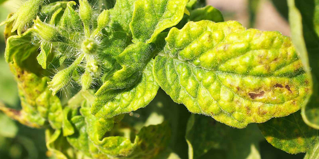
Tomato yellow leaf curl is a disease of tomato caused by Tomato yellow leaf curl virus. In March 2007, it was identified for the first time in California and currently has a limited distribution.  Infected tomato plants initially show stunted and erect or upright plant growth; plants infected at an early stage of growth will show severe stunting. However, the most diagnostic symptoms are those in leaves. Leaves of infected plants are small and curl upward; and show strong crumpling and interveinal and marginal yellowing. The internodes of infected plants become shortened and, together with the stunted growth, plants often take on a bushy appearance, which is sometimes referred to as 'bonsai' or broccoli'-like growth. Flowers formed on infected plants commonly do not develop and fall off (abscise). Fruit production is dramatically reduced, particularly when plants are infected at an early age, and it is not uncommon for losses of 100% to be experienced in fields with heavily

Tomato Mosaic Virus

Image
Tomato mosaic virus (ToMV) is a member of family tobamoviridae and belongs to the genus tobamovirus, which is a plant pathogenic virus. It is found worldwide and affects tomatoes and many other wide host range plants including many agricultural crops and weeds such as tobacco and beans, all of which can serve as inoculum sources. The tomato crop is highly susceptible to the Tomato mosaic virus (ToMV). The symptoms vary from tiles, wrinkle, reduction and curvature of leaflets, and irregular ripening of fruits. This disease requires attention because of its easy dissemination by contact, cultural practices, or contaminated seed. Tomato mosaic virus symptoms can be found at any stage of growth and all parts of the plant may be infected. ToMV causes yellow mosaic symptoms on the leaves and tomato fruits. Tomato mosaic virus symptoms are seen as a general mottling or mosaic appearance on foliage. Light and darker green mosaic leaf mottle, sometimes with distortion of younger leaves; this is

Tomato Target Spot

Image
Target spot on tomato fruit is difficult to recognize in the early stages, as the disease resembles several other fungal diseases of tomatoes. However, as diseased tomatoes ripen and turn from green to red, the fruit displays circular spots with concentric, target-like rings and a velvety black, fungal lesion in the center. The “targets” become pitted and larger as the tomato matures. Management Remove old plant debris at the end of the growing season; otherwise, the spores will travel from debris to newly planted tomatoes in the following growing season, thus beginning the disease anew. Dispose of the debris properly and don’t place it on your compost pile unless you’re sure your compost gets hot enough to kill the spores. Rotate crops and don’t plant tomatoes in areas where other disease-prone plants have been located in the past year – primarily eggplant, peppers, potatoes or, of course – tomatoes. Rutgers University Extension recommends a three-year rotation cycle to reduce soil-bo

Tomato Two Spotted Spider Mite

Image
The two-spotted spider mite is the most common mite species that attacks vegetable and fruit crops. Spider mites can occur in tomato, eggplant, potato, vine crops such as melons, cucumbers, and other crops. Two-spotted spider mites are one of the most important pests of eggplant. They have up to 20 generations per year and are favored by excess nitrogen and dry and dusty conditions. Outbreaks are often caused by the use of broad-spectrum insecticides which interfere with the numerous natural enemies that help to manage mite populations. As with most pests, catching the problem early will mean easier control. Feeding injury often gives the top leaf surfaces a mottled or speckled, dull appearance. Leaves then turn yellow and drop. Large populations produce visible webbing that can completely cover the leaves. In the field, spider mites are favored by hot dry weather, which also aggravates injury by stressing the plant. Damage is often underestimated since the wounds and the pest are not

Tomato Septoria Leaf Spot

Image
Septoria leaf spot, also called Septoria blight, is a very common disease of tomatoes.1 It is caused by a fungus (Septoria lycopersici) and can affect tomatoes and other plants in the Solanaceae family, especially potatoes and eggplant, just about anywhere in the world. Although Septoria leaf spot is not necessarily fatal for your tomato plants, it spreads rapidly and can quickly defoliate and weaken the plants, rendering them unable to bear fruit to maturity.  Septoria leaf spot is caused by a fungus, Septoria lycopersici. It is one of the most destructive diseases of tomato foliage and is particularly severe in areas where wet, humid weather persists for extended periods. Septoria leaf spot usually appears on the lower leaves after the first fruit sets. Spots are circular, about 1/16 to 1/4 inch in diameter with dark brown margins and tan to gray centers with small black fruiting structures. Characteristically, there are many spots per leaf. This disease spreads upwards from oldest

Tomato Leaf Mold

Image
Leaf mold is a common fungal disease that affects tomatoes that are cultivated in especially humid environments. Feeding off of the humidity, the mold grows and thrives after attaching itself to the leaves of the tomato plant. Once the mold is noticed, immediate action is required to save the plant, otherwise the foliage will be compromised, which will reduce the amount of tomatoes your plants will yield. Fortunately for tomato growers, this fungal disease is relatively easy to treat and cure. The first signs of leaf mold are, as you might expect, on the leaves themselves, as the topside of the leaves start to develop small gray, yellow, white, or pale green patches. The underside of the leaves begin to develop a fuzzy texture and turn purple. Oftentimes, the fuzz will appear in an olive green color. The fuzzy texture is actually the spores of the mold fungus. The fruit of the tomato plant is rarely infected by leaf mold. As the disease progresses, the infected tissue of the leaves bec

Tomato Late Blight

Image
Late blight is caused by the oomycete Phytophthora infestans. Oomycetes are fungus-like organisms also called water molds, but they are not true fungi. There are many different strains of P. infestans. These are called clonal lineages and designated by a number code (i.e. US-23). Many clonal lineages affect both tomato and potato, but some lineages are specific to one host or the other. The host range is typically limited to potato and tomato, but hairy nightshade (Solanum physalifolium) is a closely related weed that can readily become infected and may contribute to disease spread. Under ideal conditions, such as a greenhouse, petunia also may become infected. Phytophthora infestans can overwinter in Minnesota if protected in potato cull piles. Overwintering in a tomato production system is unlikely but infected tomato fruits may give rise to infected volunteer seedlings the following season. The most common routes of introduction each season are infected potato seed tubers, infected

Tomato Bacterial Spot

Image
On tomato, leaf lesions are initially circular and water-soaked and may be surrounded by a faint yellow halo. In general, spots are dark brown to black and circular on leaves and stems. Spots rarely develop to more than 3 mm in diameter. Lesions can coalesce causing a blighted appearance of leaves and a general yellowing may occur on leaves with multiple lesions. Infected pepper leaves drop prematurely; this exposes fruit to the sun and results in sun scalding. Fruit lesions begin as small, slightly raised blisters, which become dark brown, scab-like, and can appear slightly raised as they increase in size. On leaflets, bacterial spot can be easily confused with the early symptoms of bacterial speck, early blight, gray leaf spot, target spot, or Septoria leaf spot. When Xanthomonas is present, bacteria will ooze (also referred to as bacterial streaming) from infected tissue and can be observed under a light microscope. Bacterial streaming will not be observed in lesions caused by funga

Strawberry Leaf Scorch

Image
Leaf scorch is caused by the fungus Diplocarpon earliana. Symptoms of leaf scorch consist of numerous small, irregular, purplish spots or “blotches” that develop on the upper surface of leaves. The centers of the blotches become brownish. Blotches may coalesce until they nearly cover the leaflet, which then appears purplish to reddish to brown. The fungus overwinters on infected leaves. The fungus produces spore forming structures in the spring on both surfaces of dead leaves. These structures produce spores abundantly in midsummer. In the presence of free water, these spores can germinate and infect the plant within 24 hours. Older and middle-aged leaves are infected more easily than young ones. Scorched strawberry leaves are caused by a fungal infection which affects the foliage of strawberry plantings. The fungus responsible is called Diplocarpon earliana. Strawberries with leaf scorch may first show signs of issue with the development of small purplish blemishes that occur on the t

Squash Powdery Mildew

Image
  Powdery mildew, mainly caused by the fungus Podosphaera xanthii, infects all cucurbits, including muskmelons, squash, cucumbers, gourds, watermelons and pumpkins. In severe cases, powdery mildew can cause premature death of leaves, and reduce yield and fruit quality. Causes Powdery mildew infections favor humid conditions with temperatures around 68-81° F. In warm, dry conditions, new spores form and easily spread the disease. Symptoms of powdery mildew first appear mid to late summer in Minnesota.  The older leaves are more susceptible and powdery mildew will infect them first. Wind blows spores produced in leaf spots to infect other leaves. Under favorable conditions, powdery mildew can spread very rapidly, often covering all of the leaves. Although powdery mildew primarily infects leaves and vines, infections occasionally occur on cucumber or melon fruit. Powdery mildew does not directly infect squash fruit. Fewer and smaller fruit grow on infected plants. Reduced fruit quality oc

Potato Late Blight

Image
Found on tomato and potato plants, late blight is caused by the fungus Phytophthora infestans and is common throughout the United States. True to its name, the disease occurs later in the growing season with symptoms often not appearing until after blossom. Late blight first appears on the lower, older leaves as water-soaked, gray-green spots. As the disease matures, these spots darken and a white fungal growth forms on the undersides. Eventually the entire plant will become infected. Crops can be severely damaged Unlike other fungal diseases, this plant problem does not overwinter in the soil or on garden trash. Instead the spores are introduced by infected tubers, transplants or seeds. Wind will also carry the disease from nearby gardens. Warm temperatures (70-80˚F) and wet, humid conditions promote its rapid spread. Note: Late Blight was responsible for the Irish potato famine (1845-1849).  Treatment Destroy all cull and volunteer potatoes. Plant late blight-free seed tubers. Do not

Potato Early Blight

Image
  Early blight of potato is a common disease found in most potato growing regions. The disease is caused by the fungus Alternaria solani, which can also afflict tomatoes and other members of the potato family. Potatoes become infected with early blight when foliage has become excessively wet due to rain, fog, dew, or irrigation. Although not a terminal disease, severe infections can be fairly detrimental. In contrast to its name, early blight rarely develops early; it actually usually affects mature foliage rather than young, tender leaves. Symptoms Early blight rarely affects young plants. Symptoms first occur on the lower or oldest leaves of the plant. Dark, brown spots appear on this older foliage and, as the disease progresses, enlarge, taking on an angular shape. These lesions often look like a target and, in fact, the disease is sometimes referred to as target spot. As the spots enlarge, they may cause the entire leaf to yellow and die, but remain on the plant. Dark brown to blac

Bell Pepper Bacterial Spot

Image
Leaf spots that appear on the lower surface of older leaves as small, pimples and on the upper leaf surface as small water-soaked spots are a symptom of bacterial spot. This is an important pepper disease in Maryland. It also occasionally attacks tomatoes. Eventually, the spots develop gray to tan centers with darker borders. Lesions enlarge during warm, humid weather. Leaves may then turn yellow, then brown and drop. Lesions may also develop on stems. Fruits develop small, raised rough spots that do not affect eating quality. Severely infected leaves will drop resulting in sun-scald of peppers. Bacterial leaf spot is spread by splashing rain and working with wet, infected plants. This disease can defoliate plants during wet weather. Hot, dry weather slows the spread of this disease. The disease can come in on seed or transplants and can overwinter in crop residue and soil. Management You can minimize problems with bacterial spot by following these tips: Select resistant varieties Purc

Peach Bacterial Spot

Image
Bacterial leaf spot of peach, also known as bacterial shot hole, is a common disease on older peach trees and nectarines. This peach tree leaf spot disease is caused by the bacterium Xanthomonas campestris pv. pruni. Bacterial spot on peach trees results in loss of fruit and the overall malaise of trees caused by recurrent defoliation. Also, these weakened trees are more susceptible to winter injury. The most characteristic sign of peach tree leaf spot are angular purple to purple-brown spots on foliage, followed by the center of lesion falling out, giving the leaves a “shot hole” appearance. Leaves soon turn yellow and drop. Fruit has small water-soaked markings that enlarge and merge to eventually cover large areas. Cracking or pitting occurs along the lesions as the fruit grows, enabling brown rot fungus to infiltrate the fruit. Bacterial leaf spot also affects current season growth. Two types of cankers can be spotted on twigs. “Summer cankers” appear on green twigs after leaf spot

Orange Huanglongbing

Image
Huanglongbing (HLB) or citrus greening is the most severe citrus disease, currently devastating the citrus industry worldwide. The presumed causal bacterial agent Candidatus Liberibacter spp. affects tree health as well as fruit development, ripening and quality of citrus fruits and juice. Fruit from infected orange trees can be either symptomatic or asymptomatic. Symptomatic oranges are small, asymmetrical and greener than healthy fruit. Furthermore, symptomatic oranges show higher titratable acidity and lower soluble solids, solids/acids ratio, total sugars, and malic acid levels. Among flavor volatiles, ethyl butanoate, valencene, decanal and other ethyl esters are lower, but many monoterpenes are higher in symptomatic fruit compared to healthy and asymptomatic fruit. The disease also causes an increase in secondary metabolites in the orange peel and pulp, including hydroxycinnamic acids, limonin, nomilin, narirutin, and hesperidin. Resulting from these chemical changes, juice made

Grape Leaf Blight

Image
  The yellow-green disease spots gradually appear on the fronts of the grape leaves with downy mildew, and white frosty mildew appears on the backs of the leaves. Leaf blight produces dark brown patches on the surface of grape leaves

Grape Black Measles

Image
Grapevine measles, also called esca, black measles or Spanish measles, has long plagued grape growers with its cryptic expression of symptoms and, for a long time, a lack of identifiable causal organism(s). The name ‘measles’ refers to the superficial spots found on the fruit (Fig. 1). During the season, the spots may coalesce over the skin surface, making berries black in appearance. Spotting can develop anytime between fruit set and a few days prior to harvest. Berries affected at fruit set tend not to mature and will shrivel and dry up. In addition to spotting, fruit affected later in the season will also have an acrid taste. Leaf symptoms are characterized by a ‘tiger stripe’ pattern (Fig 2-bottom leaf) when infections are severe from year to year. Mild infections can produce leaf symptoms (Fig. 2-upper leaf) that can be confused with other diseases or nutritional deficiencies. White cultivars will display areas of chlorosis followed by necrosis, while red cultivars are characteriz

Grape Black Rot

Image
Black rot, caused by the fungus Guignardia bidwellii, is a serious disease of cultivated and wild grapes. The disease is most destructive in warm, wet seasons. It attacks all green parts of the vine – leaves, shoots, leaf and fruit stems, tendrils, and fruit. The most damaging effect is to the fruit. Note: Guignardia bidwellii forma parthenocissi causes a leaf spot on Boston ivy and Virginia creeper. Control is as for black rot of grapes. Warm, muggy weather in the spring and summer, along with unsprayed fruit of susceptible varieties, may cause fruit to become almost completely rotted by harvest time. Black rot is not difficult to control if good cultural practices are followed along with the use of protective fungicide sprays. Symptoms and Diagnosis Leaves: Reddish brown and circular to angular spots appear on the upper surface of the leaves starting in late spring. As spots merge, they form irregular, reddish brown blotches. The number of spots or lesions per leaf varies from 2 to m

Corn Northern Leaf Blight

Image
  Northern corn leaf blight (NCLB) is caused by the fungus Setosphaeria turcica. Symptoms usually appear first on the lower leaves. Leaf lesions are long (1 to 6 inches) and elliptical, gray-green at first but then turn pale gray or tan. Under moist conditions, dark gray spores are produced, usually on the lower leaf surface, which give lesions a "dirty" gray appearance. Entire leaves on severely blighted plants can die, so individual lesions are not visible. Lesions may occur on the outer husk of ears, but the kernels are not infected. On hybrids that contain an Ht gene for resistance to the fungus, lesions are smaller, chlorotic, and may develop into linear streaks. These lesions rarely produce spores. Lesions can eventually expand to a more oblong or “cigar” shape. They may also coalesce to form large areas of necrotic tissue. Dark spores may be observed in the lesions with the aid of a hand lens. Symptoms often begin on lower leaves; spores produced by primary lesions are

Corn Common Rust

Image
  Common corn rust, caused by the fungus Puccinia sorghi, is the most frequently occurring of the two primary rust diseases of corn in the U.S., but it rarely causes significant yield losses in Ohio field (dent) corn. Occasionally field corn, particularly in the southern half of the state, does become severely affected when weather conditions favor the development and spread of the rust fungus. Sweet corn is generally more susceptible than field corn. In years with exceptionally cool summers, and especially on late-planted fields or sweet corn, yield losses may occur when the leaves at and above the ears become severely diseased before grain fill is complete. Symptoms Although a few rust pustules can always be found in corn fields throughout the growing season, symptoms generally do not appear until after tasseling. These can be easily recognized and distinguished from other diseases by the development of dark, reddish-brown pustules (uredinia) scattered over both the upper and lower s

Corn Gray Leaf Spot

Image
Gray leaf spot, caused by the fungus Cercospora zeae-maydis, occurs virtually every growing season. If conditions favor disease development, economic losses can occur. Symptoms first appear on lower leaves about two to three weeks before tasseling. The leaf lesions are long (up to 2 inches), narrow, rectangular, and light tan colored. Later, the lesions can turn gray. They are usually delimited by leaf veins but can join together and kill entire leaves. All corn hybrids and inbreds are susceptible to Gray Leaf Spot (GLS). In minimum tillage fields with a history of GLS, symptoms can occur on the lower leaves prior to tasseling. Immature GLS lesions can be easily confused with lesions caused by other pathogens; e.g., eyespot (Kabatiella), anthracnose (Colletrichum). Mature lesions on leaves are rectangular in shape and restricted by leaf veins; reverse lighting reveals a yellow halo on most hybrids. Mature lesions are diagnostic and easily distinguishable from other diseases. As lesions